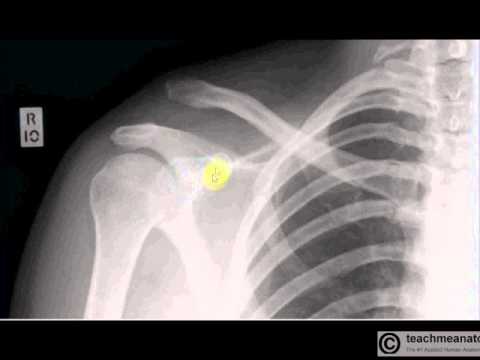
A video tutorial in interpreting radiographs of the shoulder joint and surrounding areas. This is the second video in a series of five by TeachMeAnatomy — have a look at the rest at www.teachmeanatomy.info
This video covers the following:
– The normal radiographic anatomy of the shoulder joint
– Surgical neck fractures and complications
– Acromioclavicular dislocation
– Clavicular fractures
– Anterior and posterior shoulder dislocations

No comments:
Post a Comment