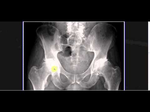
A video tutorial in interpreting radiographs of the pelvis, hip joint and femur. This is the fourth video in a series of five by TeachMeAnatomy — have a look at the rest at www.teachmeanatomy.info.
This video covers the following:
– Normal radiographic anatomy of the pelvis, hip joint and femur
– Different between male and female pelvic anatomy
– Osteoarthritis of the hip joint
– Fractures of the femoral neck
– Differentiating between intracapsular and extracapsular fractures
– Posterior hip dislocations
– Fractures of the femoral shaft

No comments:
Post a Comment